Blue tongue in dogs
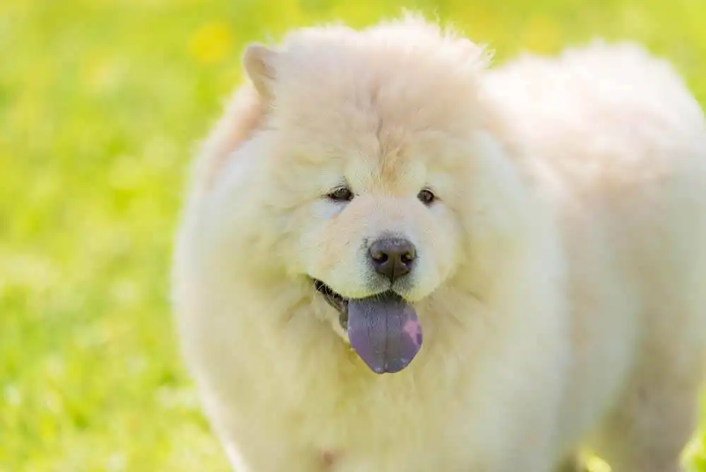
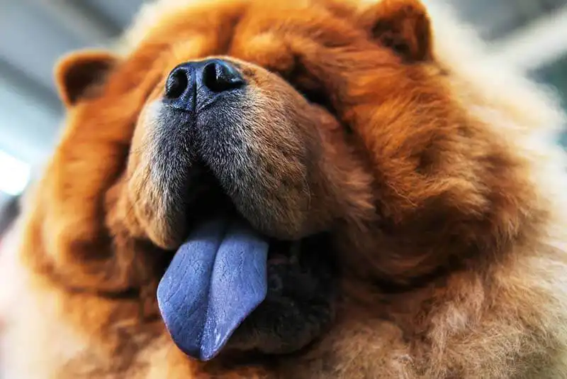
A blue tongue in a dog does not always indicate health problems. For some breeds, such as the Chow Chow, a blue tongue is considered normal. If the changes appear suddenly over a short period of time, it makes sense to see a doctor and get examined. They may indicate serious health problems.
About cyanosis
Oxygenated blood is bright red, so the tongue should normally be pale pink to pink.
Altered, deoxygenated blood is blue or brown in color, so a blue or purple tongue and the inner surface of the cheeks and gums indicate an acute manifestation of oxygen deficiency of any origin in the pet.
Types of cyanosis
In false cyanosis, the bluish tint is observed due to dyes entering the blood or the surface of the tongue, which are deposited in the skin and mucous membranes.
True cyanosis is a manifestation of cardiac or respiratory failure, characterized by the accumulation in the blood of a large amount of hemoglobin that is not saturated with oxygen.
In central cyanosis, cyanosis occurs as a result of disruptions in the functioning of the central circulatory system. Its occurrence is due to a significant decrease in the level of oxygen in the body’s blood – cyanosis appears on the skin, mucous membranes of the oral cavity, conjunctiva of the eye (mucous membrane), etc.
Peripheral cyanosis is a disorder that is specific to one organ or one part of the body. This could be an injured limb or an organ with a disrupted blood supply.
Why Does a Dog Have a Blue Tongue – 10 Reasons
Norm for some breeds
Pigmented mucous membrane can be normal for almost any breed, but this manifestation is most often found in Chow Chows and Shar Peis . In this case, this coloring is observed in the dog throughout its life.
Narrowing of the tracheal lumen or tracheal collapse
This pathology has many causes – from congenital predisposition to acute allergic reaction. It causes a violation of the animal’s respiratory ability – inhalations become short and unproductive, alternating with coughing. This provokes the development of general respiratory failure and blue tongue.
Violation of the integrity of the respiratory tract
Trauma to the trachea, larynx, lungs, neoplasms can lead to the appearance of cyanosis of the tongue. Trauma to the airways or lung tissue itself implies a violation of the dog’s ability to normally inhale and exhale.
Respiratory muscle failure
Breathing is carried out by a number of respiratory muscles. In case of excessive relaxation of skeletal muscles, disruption of the nerve fibers or the respiratory center that sends impulses, oxygen starvation occurs, which is manifested by a blue tongue.
A buildup of air or fluid in the chest
Air or fluid in the chest does not allow the lungs to properly expand and fill with blood, which in itself prevents the blood from being saturated with oxygen. As a result, oxygen starvation occurs.
Pulmonary edema of any origin
The fluid filling the lungs disrupts their functioning and, accordingly, causes a number of symptoms of oxygen starvation. Including the dog’s tongue turning blue.
Heart pathologies
Various pathologies such as malfunction of the valve system, the presence of congenital anomalies, inflammation of the heart muscle, tumor process, cardiac parasites – all this disrupts the heart’s flow system. There is stagnation of blood in the pulmonary circulation, which prevents normal saturation of blood in the lungs with oxygen.
Elongation of the soft palate – brachycephalic syndrome
This syndrome is typical for short-muzzled dogs – pugs , French and English bulldogs , etc. One of its signs is thickening and lengthening of the soft palate. This soft structure blocks the lumen of the dog’s larynx and prevents it from taking a normal breath. During periods of exacerbation of respiratory failure, it can thicken so much that it does not allow the animal to take a breath at all. In this regard, manifestations of respiratory failure can be observed.
Bronchitis
An allergic reaction, an autoimmune process (excessively increased immunity), viral diseases, fungal infections of the lower respiratory tract cause spasm of the bronchial tissue. It is characterized by respiratory failure and a blue tongue in dogs.
Eating pigmented foods
Some products and substances contain a pigment that can stain the skin and mucous membranes of the oral cavity. In particular, the dog’s tongue can turn blue, brown, purple, violet. These include blueberries, mulberries, beets, activated carbon.
Associated symptoms
In case of bronchitis, tracheal collapse, brachycephalic syndrome, and injuries, the following may additionally be observed: coughing, coughing up mucus or blood clots, and reverse sneezing syndrome.
For pulmonary edema, prolonged oxygen starvation, a tense posture of the sphinx is characteristic, in which the animal lies on its stomach, its sides become sunken. The dog makes significant efforts to perform the act of inhalation. It may also experience a decrease in body temperature.
With all types of oxygen starvation, the following are observed: dyspnea of a mixed type (both on inhalation and exhalation), bluish visible mucous membranes (oral mucosa, tongue, conjunctiva of the eye), non-pigmented nasal mirror and skin, frequent shallow breathing.
In false cyanosis, the tongue gradually loses its strange color after rinsing the mouth with water or contact with other food.
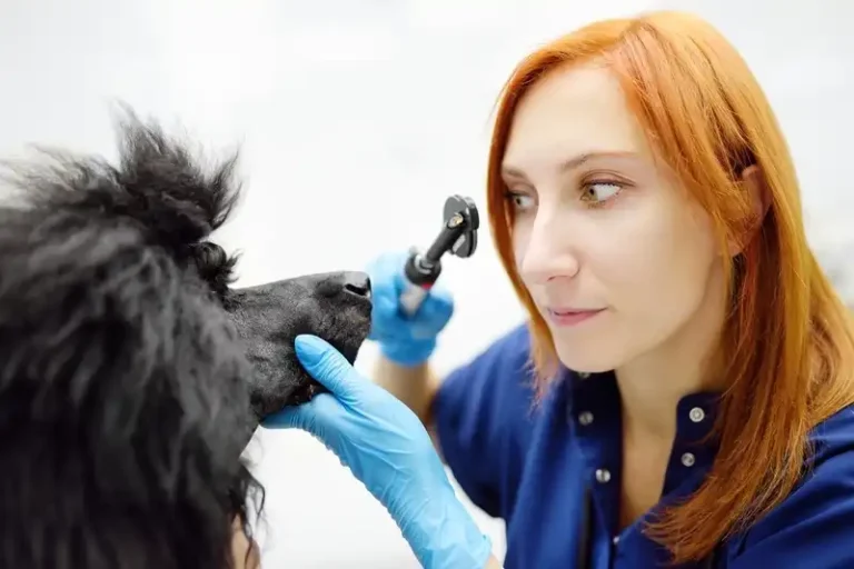
Diagnostics
- For any type of pathology, the following will initially be prescribed:
- X-ray diagnostics of the chest and neck. It is performed in a straight and two lateral positions – right and left
- Chest ultrasound – a short T-Fast protocol to exclude or confirm acute respiratory or cardiogenic (extreme left ventricular failure) diseases
- General clinical and biochemical blood tests
- If there is fluid in the chest, a cytological (microscopic examination of one type of cell) and biochemical examination of the fluid is additionally performed.
- If there is a suspicion of a neoplasm in the chest or upper respiratory tract, the following is prescribed:
- Chest computed tomography
Histological (analysis of the structure of cells and tissues) and cytological examination of the formation, taken during diagnostic thoracotomy (examination of the chest cavity) or endoscopic examination
If a diaphragmatic hernia is suspected, an X-ray examination with contrast (using a contrast agent) will be required.
In case of pulmonary edema, the doctor prescribes an ultrasound and ECG of the heart. This is necessary to confirm or exclude the cardiogenic origin of this pathology.
Bronchitis, asthma, tracheal collapse require bronchoalveolar lavage. During this procedure, a sterile saline solution is introduced into the lumen of the respiratory tract of a sedated (immobilized) animal, which is then drained back. This fluid is sent for a comprehensive examination: PCR for respiratory infections, cytological examination, and culture to detect sensitivity to antibiotics.
Also, for these diseases, tracheoscopy and bronchoscopy are prescribed – endoscopic examination of the respiratory tract.

Treatment
Treatment is provided only after the animal’s condition has stabilized and primary diagnostic data has been obtained – X-ray, ultrasound, blood tests.
Primary therapy for any disease is aimed at stabilizing the animal’s condition. It includes:
Oxygen therapy is a method that helps increase the amount of oxygen in the air an animal inhales.
Sedative therapy. Often it is necessary to take sedatives (calming) such as tranquilizers/anticonvulsants (trazadone, gabapentin, vet-uspokoin) to even out breathing.
Monitoring temperature and pressure, glucose levels, and correcting them if necessary.
Free fluid or air in the chest requires immediate removal. To do this, the hair is cut off, the skin surface is treated and by puncturing the soft tissues in the intercostal space, a needle is inserted into the chest, through which air or fluid is removed with syringes, creating negative pressure.
If necessary, active drainage is installed – a permanently installed tube. A bulb is attached to it, which builds up pressure and constantly helps remove air or fluid from the chest.
In case of active loss of protein with breast fluid, it may be necessary to replenish its level by artificial intravenous administration of pure albumin, plasma or blood of another animal.
In case of blood loss, serious injuries, tumor processes, it is necessary:
blood transfusion under the supervision of a doctor strictly in a veterinary clinic
surgical intervention – removal of formations, surgical treatment of injuries, diaphragmatic hernia, etc.
installation of a tracheostomy – a tube that forms an airway through the trachea. It is used in cases of significant injuries to the larynx, neck, and head.
Cardiogenic pulmonary edema requires diuretic therapy with various drugs (Furosemide, Torasemide, Apcard, Veroshpiron, and others), as well as the use of drugs that correct blood pressure (Dopamine, Dobutamine). The doctor may also prescribe Vetmedin to stimulate cardiac output.
Tracheal collapse, bronchitis, bronchopneumonia require hormonal therapy in the form of inhalation or oral administration (through the mouth) of Prednisolone, Dexamethasone, Budesonide, bronchodilators (Salbutamol) or antibacterial drugs (Baytril).
Providing first aid
Unfortunately, it is impossible to provide quality first aid to a pet with a blue or already burgundy tongue at home. A blue tongue in a dog that is also breathing heavily is usually an emergency. Therefore, if such a change is detected in combination with heavy breathing, lethargy or excessive excitability, it is necessary to immediately transport the animal to the clinic for examination and first aid. During transportation, it is important to put the pet in a comfortable position – on its stomach. It should also be provided with a large amount of freely flowing air or oxygen (oxygen cylinders can be used).
Prevention
Annual medical examination allows to detect most diseases, deterioration of the condition, up to emergency. Under the supervision of a specialized physician, it will be possible to prevent pulmonary edema, bronchial asthma, etc.
The occurrence of brachycephalic syndrome can be prevented by timely plastic surgery of the nasal mirror in a short-muzzled dog. It is better to perform the operation at an early age. Injuries, allergic reactions, neurological disorders cannot be predicted. These conditions themselves require immediate intervention of a veterinarian.
Blue Tongue in Dogs: Summary
Blueness of the tongue or oral mucosa does not always indicate the presence of diseases in the animal. Some breeds have a blue tongue by nature or acquire it by eating colored foods.
With cyanosis, the pathological blueness of the tongue indicates a lack of oxygen in the animal’s body and an oversaturation with carbon dioxide – it suffocates.
The main reasons why a dog may have a blue tongue are: tracheal collapse, trauma, respiratory muscle failure, fluid or air accumulation in the chest, pulmonary edema, heart disease, elongation of the soft palate, bronchitis or bronchopneumonia.
Basic diagnostics include: X-ray, chest ultrasound, heart ultrasound, ECG, tracheoscopy and bronchoscopy, etc.
Treatment of this condition directly depends on the diagnosis. What unites all diseases is the urgency of the condition – immediate medical intervention and stabilization in a hospital setting are required.
First aid at home for a dog with a burgundy or bluish tongue is impossible. The owner must immediately transport the pet to a veterinary clinic.
The main prevention of this condition is annual medical examination and observation by a veterinarian of an animal with chronic diseases.